This is the content of the pop-over!


DANB's NYR (NYR) Practice Tests & Test Prep by Exam Edge - Exam Info


** Sample images, content may not apply to your exam **


The more you know about the DANB's NYR exam the better prepared you will be! Our practice tests are designed to help you master both the subject matter and the art of test-taking to be sure you are fully prepared for your exam.

Here are a few things to think about:

  • What is the DANB's NYR certification exam?
  • Who is Responsible for the DANB exam?
  • Am I eligibility for the DANB's NYR Exam?
  • What is the best way to ensure your succes on the first try?
  • The benefits of using Exam Edge to pass your DANB's NYR exam.

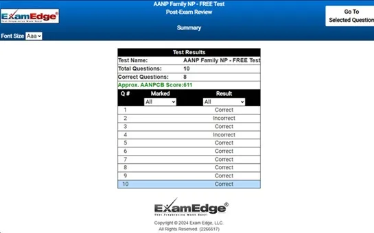
Not ready to purchase our complete practice tests yet? Start with a DANB's New York Radiology FREE Practice Test first!

Excellent


Select Quantity

Buy one or save big with a Multi Test Value Pack for the DANB's NYR exam.

All transactions
secured and encrypted
All prices are
in US dollars
Get Instant Online Access Now!


Additional test information
Back To General Exam Info

DANB's New York Radiology - Additional Information

At ExamEdge.com, we focus on making our clients' career dreams come true by offering world-class practice tests designed to cover the same topics and content areas tested on the actual Dental Assisting National Board DANB's New York Radiology (NYR) Certification Exam. Our comprehensive DANB's New York Radiology practice tests are designed to mimic the actual exam. You will gain an understanding of the types of questions and information you will encounter when you take your Dental Assisting National Board DANB's New York Radiology Certification Exam. Our DANB's New York Radiology Practice Tests allow you to review your answers and identify areas of improvement so you will be fully prepared for the upcoming exam and walk out of the test feeling confident in your results.

Because our practice tests are web-based, there is no software to install and no need to wait for a shipment to arrive to start studying. Your DANB's New York Radiology practice tests are available to you anytime from anywhere on any device, allowing you to study when it works best for you. There are 15 practice tests available, each with 100 questions and detailed explanations to help you study. Every exam is designed to cover all of the aspects of the DANB's NYR exam, ensuring you have the knowledge you need to be successful!


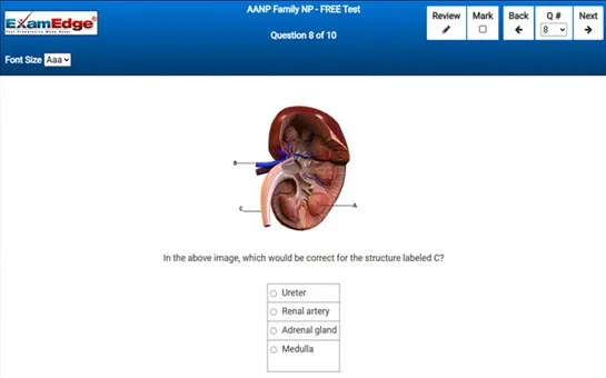
DANB's New York Radiology - Additional Info Sample Questions

Which of the following indicates that you are viewing the mandibular molars?


Correct Answer:
two divergent roots.


in dental anatomy and radiography, differentiating between the types of teeth, especially molars, is crucial for accurate diagnosis and treatment planning. the question at hand involves identifying features that indicate a view of the mandibular molars on a radiograph.

the correct answer to the question is "two divergent roots." this choice is accurate because mandibular molars typically exhibit two roots that diverge from each other. these roots are known as the mesial and distal roots. the mesial root is located towards the midline of the face, and the distal root is positioned towards the back of the mouth. this configuration is distinct and helps in distinguishing mandibular molars from other teeth, such as maxillary molars, which generally have three roots.

the other options provided in the question can be analyzed to understand why they do not correctly describe the mandibular molars: - "three roots": this is a characteristic of maxillary molars, not mandibular molars. maxillary molars usually have three roots, two buccal (toward the cheek) and one palatal (toward the palate), which helps in supporting the tooth in the denser maxillary bone. - "very large crowns": while mandibular molars do have relatively large crowns compared to other teeth, this feature alone is not specific enough to distinguish them on a radiograph, as maxillary molars also have large crowns. - "large radiolucent areas": radiolucent areas on a dental radiograph appear dark and indicate regions where radiation passes through with less resistance, such as cavities or soft tissues. this description does not specifically pertain to the identification of mandibular molars.

understanding the root configuration on dental radiographs is fundamental for proper identification and orientation of the images. this knowledge assists dental professionals in diagnosing diseases such as decay and infection localized to specific roots, planning extractions, and other dental procedures that require precise anatomical knowledge. hence, recognizing the two divergent roots of mandibular molars is key in the context of dental radiography.