Understanding the exact breakdown of the ABIM Cardiovascular Disease test will help you know what to expect and how to most effectively prepare.
The ABIM Cardiovascular Disease has 200 multiple-choice questions
.
The exam will be broken down into the sections below:
ABIM Cardiovascular Disease Exam Blueprint
Domain Name
%
Number of Questions
Arrhythmias
15%
30
Coronary Artery Disease
21.5%
43
Heart Failure and Cardiomyopathy
17%
34
Valvular Disease
15%
30
Pericardial Disease
4%
8
Congenital Heart Disease
5%
10
Vascular Diseases
6%
12
Systemic Hypertension and Hypotension
7.5%
15
Pulmonary Circulation Disorders
3%
6
Systemic Disorders Affecting the Circulatory System
4%
8
Normal Cardiovascular Anatomy and Physiology
2%
4
ABIM Cardiovascular Disease Study Tips by Domain
When a tachyarrhythmia is present, first decide if the patient is hemodynamically unstable (hypotension, ischemic chest pain, altered mental status, shock, acute HF)—this is a priority rule for immediate synchronized cardioversion rather than additional diagnostic maneuvers.
For regular narrow-complex SVT, vagal maneuvers then adenosine is first-line, but a key contraindication/red flag is irregular wide-complex tachycardia (suspected preexcited AF) where AV nodal blockers can precipitate VF.
Atrial fibrillation management hinges on stroke prevention: use CHA2DS2-VASc to guide anticoagulation, and don’t fall for the trap that restoration of sinus rhythm eliminates the need for anticoagulation when risk is high.
In wide-complex tachycardia, assume ventricular tachycardia until proven otherwise—a common ABIM trap is treating presumed SVT with aberrancy using AV nodal blockers in an older patient with structural heart disease.
Bradyarrhythmias: symptomatic bradycardia or high-grade AV block requires atropine and often temporary pacing, and a red flag is Mobitz II or complete heart block (especially with wide QRS) where permanent pacing is typically indicated.
QT prolongation with polymorphic VT (torsades de pointes) is treated with IV magnesium and correction of K/Mg, and the practical cue is to stop QT-prolonging drugs and avoid amiodarone/sotalol when acquired long-QT is the driver.
In suspected ACS, obtain serial ECGs and high-sensitivity troponins; red flag: new ST-elevation or posterior MI pattern warrants immediate reperfusion rather than waiting for biomarkers.
Use DAPT after ACS/PCI but reassess bleeding risk; common trap: continuing prasugrel in prior stroke/TIA (contraindicated) or forgetting GI bleed history when choosing P2Y12 inhibitor.
For stable chest pain, match testing to pretest probability and ability to exercise; priority rule: avoid routine stress testing in very low-risk patients (false positives lead to unnecessary angiography).
In STEMI, choose primary PCI when feasible; threshold cue: if first medical contact-to-device time will exceed guideline targets, give fibrinolysis unless contraindicated (e.g., prior intracranial hemorrhage).
High-intensity statin is foundational in clinical ASCVD; common trap: stopping statin for mild, asymptomatic transaminase elevation—evaluate other causes and continue unless true hepatotoxicity or rhabdomyolysis.
Recognize mechanical complications post-MI (papillary muscle rupture, VSD, free wall rupture); red flag: new harsh murmur, pulmonary edema, or sudden hypotension 2–7 days after MI requires urgent echo and surgical consultation.
Severe aortic stenosis is high-risk when Vmax =4.0 m/s, mean gradient =40 mmHg, or AVA =1.0 cm2; red flag is syncope, angina, or dyspnea — don’t delay valve intervention once symptoms appear.
In chronic aortic regurgitation, operate when symptoms develop or when LVEF =55% or LVESD >50 mm (or >25 mm/m2); common trap is being reassured by a “normal” EF in a markedly dilated LV.
Primary (degenerative) MR: refer for repair when symptomatic severe MR or when LVEF =60% and/or LVESD =40 mm even if asymptomatic; red flag is new atrial fibrillation or pulmonary hypertension (PASP >50 mmHg at rest).
Secondary (functional) MR: priority is GDMT for HFrEF and CRT when indicated; common trap is sending all severe MR to surgery — consider transcatheter edge-to-edge repair only if symptoms persist and anatomy is suitable.
Mitral stenosis severity: valve area =1.5 cm2 is significant and =1.0 cm2 is severe; red flag is atrial fibrillation or prior embolic event — anticoagulate with warfarin (not DOACs) in rheumatic MS with AF.
Suspected infective endocarditis in valvular disease: obtain 3 sets of blood cultures before antibiotics unless unstable; common trap is relying on TTE alone — TEE is preferred for prosthetic valves or persistent bacteremia.
Severe aortic stenosis is high-risk when Vmax =4.0 m/s, mean gradient =40 mmHg, or AVA =1.0 cm2; red flag is syncope, angina, or dyspnea — don’t delay valve intervention once symptoms appear.
In chronic aortic regurgitation, operate when symptoms develop or when LVEF =55% or LVESD >50 mm (or >25 mm/m2); common trap is being reassured by a “normal” EF in a markedly dilated LV.
Primary (degenerative) MR: refer for repair when symptomatic severe MR or when LVEF =60% and/or LVESD =40 mm even if asymptomatic; red flag is new atrial fibrillation or pulmonary hypertension (PASP >50 mmHg at rest).
Secondary (functional) MR: priority is GDMT for HFrEF and CRT when indicated; common trap is sending all severe MR to surgery — consider transcatheter edge-to-edge repair only if symptoms persist and anatomy is suitable.
Mitral stenosis severity: valve area =1.5 cm2 is significant and =1.0 cm2 is severe; red flag is atrial fibrillation or prior embolic event — anticoagulate with warfarin (not DOACs) in rheumatic MS with AF.
Suspected infective endocarditis in valvular disease: obtain 3 sets of blood cultures before antibiotics unless unstable; common trap is relying on TTE alone — TEE is preferred for prosthetic valves or persistent bacteremia.
Acute pericarditis is diagnosed when =2 of: pleuritic chest pain, pericardial friction rub, diffuse ST elevation/PR depression, new/worsening effusion; red flag for myopericarditis is elevated troponin with reduced LV function (manage as myocarditis with activity restriction).
First-line therapy for idiopathic/viral pericarditis is NSAID (e.g., ibuprofen) plus colchicine to reduce recurrence; common trap is giving corticosteroids up front — reserve for autoimmune/uremic/pericarditis refractory to NSAID/colchicine or when NSAIDs contraindicated.
Cardiac tamponade is suggested by hypotension, elevated JVP, tachycardia, pulsus paradoxus >10 mm Hg, and echo RA/RV diastolic collapse; priority rule is urgent pericardiocentesis for hemodynamic compromise rather than waiting on further imaging.
In suspected aortic dissection (sudden tearing pain, pulse deficit, widened mediastinum), avoid anticoagulation/thrombolysis for presumed MI — this is a lethal trap; obtain emergent CTA/TEE and control BP/HR first.
Constrictive pericarditis clues include right-sided failure with Kussmaul sign, pericardial knock, and ventricular interdependence (discordant LV/RV systolic pressures with respiration); key differentiator from restrictive cardiomyopathy is respiratory variation in mitral inflow and hepatic vein diastolic flow reversal.
Pericardial effusions require etiology-based workup (malignancy, TB, uremia, hypothyroidism) and monitoring for progression; red flag for intervention is large effusion with tamponade physiology or suspected purulent/neoplastic effusion needing diagnostic drainage.
Cyanotic newborn with shock/collapse — suspect ductal-dependent CHD and start prostaglandin E1 immediately; red flag is giving only oxygen/fluids while delaying PGE1.
Fixed split S2 suggests atrial septal defect; trap is attributing a “split” that varies with respiration to ASD rather than normal physiology.
Repaired tetralogy of Fallot with severe pulmonic regurgitation can present years later with RV dilation and ventricular arrhythmias; priority cue is progressive QRS widening (e.g., >180 ms) prompting evaluation for pulmonary valve replacement/EP risk stratification.
Coarctation of the aorta — compare upper vs lower extremity BPs and femoral pulses; red flag is refractory hypertension in a young patient or arm–leg systolic gradient =20 mmHg.
Bicuspid aortic valve — screen for ascending aortopathy and counsel on family screening; trap is focusing only on the valve gradient and missing progressive aortic dilation needing serial imaging.
Eisenmenger physiology (unrepaired shunt with PAH) — avoid closing the defect once pulmonary vascular disease is established; contraindication cue is right-to-left shunting with elevated PVR where closure can precipitate RV failure.
Acute limb ischemia is a time-critical diagnosis—the red flag is the sudden onset of pain with a pulseless, cool extremity; start IV heparin immediately unless contraindicated and obtain urgent vascular surgery/endovascular consultation.
Suspected aortic dissection: treat first by lowering shear stress (IV beta-blocker before vasodilator)—common trap is giving hydralazine/nitroprusside without rate control, which can worsen dissection.
Abdominal aortic aneurysm screening is ultrasound in men age 65–75 who have ever smoked; red flag for rupture is hypotension with back/abdominal pain and a pulsatile mass—do not delay definitive imaging/OR for extensive workup.
Carotid disease: the priority rule is that symptomatic high-grade stenosis (typically 70%–99%) benefits from carotid endarterectomy if perioperative stroke/death risk is acceptable; common trap is intervening on asymptomatic moderate stenosis without optimized medical therapy.
Venous thromboembolism: for suspected pulmonary embolism with hemodynamic instability, give immediate anticoagulation and consider systemic thrombolysis if no major bleeding risk; red flag contraindication is prior intracranial hemorrhage or active bleeding.
Peripheral artery disease management prioritizes high-intensity statin and antiplatelet therapy plus supervised exercise for claudication; common trap is using compression stockings in suspected critical limb ischemia (rest pain/ulcer/gangrene) instead of urgent revascularization evaluation.
Diagnose true hypertension with proper technique (seated, correct cuff, multiple readings) and confirm with home/ambulatory BP when white-coat or masked HTN is suspected—trap: escalating meds based on a single in-office reading.
In hypertensive emergency, treat end-organ injury and lower MAP by ~20%–25% in the first hour (then to ~160/100 in 2–6 hours for most)—red flag: precipitous drops causing stroke/AKI.
Evaluate secondary causes when onset is abrupt/early, resistant (uncontrolled on 3 meds incl. diuretic), or with clues (hypokalemia, renal bruit, episodic spells)—priority rule: screen for primary aldosteronism with aldosterone–renin ratio when unexplained hypokalemia or resistant HTN.
Choose therapy to comorbidity and avoid contraindications—cue: ACEi/ARB contraindicated in pregnancy and bilateral renal artery stenosis; thiazides often worsen hyponatremia and gout.
Orthostatic hypotension is a sustained fall of =20 mmHg systolic or =10 mmHg diastolic within 3 minutes of standing—common trap: measuring too late or missing medication causes (alpha-blockers, nitrates, diuretics).
In shock or symptomatic hypotension, prioritize rapid assessment of perfusion (mental status, urine output, lactate) and treat the cause—red flag: giving beta-blockers/vasodilators before stabilizing volume/vasopressors in acute decompensation.
Suspect massive PE when hypotension/shock accompanies acute dyspnea with RV strain; priority rule: start empiric anticoagulation if pretest probability is high and bleeding risk acceptable while definitive imaging is arranged.
Use hemodynamics to separate etiologies of pulmonary hypertension (PH): post-capillary PH has PCWP =15 mmHg; common trap: labeling PH as “primary” without right-heart catheterization confirmation.
In chronic thromboembolic pulmonary hypertension (CTEPH), persistent dyspnea =3 months after PE is a red flag; priority test: V/Q scan (more sensitive than CT angiography for CTEPH screening).
In acute PE, avoid overreliance on D-dimer in high-risk patients; practical cue: use age-adjusted D-dimer (age × 10 ng/mL FEU for >50) only in low/intermediate pretest probability.
Treat pulmonary arterial hypertension (PAH) with vasoreactivity testing only in idiopathic/heritable/drug-induced PAH; contraindication/trap: do not use calcium channel blockers without a positive vasoreactivity test.
For acute RV failure from PH/PE, maintain RV perfusion and reduce afterload; red flag: aggressive diuresis or excessive PEEP can drop preload and worsen shock.
Suspect infective endocarditis in persistent bacteremia (especially Staphylococcus aureus) or new murmur; ABIM trap: treating with antibiotics before obtaining at least 2–3 sets of blood cultures unless the patient is unstable.
In atrial fibrillation with rheumatic mitral stenosis or a mechanical valve, anticoagulate with warfarin (not a DOAC); red flag is an inappropriately chosen DOAC in “valvular AF.”
For antiphospholipid syndrome with arterial thrombosis, use warfarin with an INR target typically 2.0–3.0 (higher in selected recurrent cases); common trap is relying on a positive lupus anticoagulant to mean “bleeding risk” rather than thrombosis.
Recognize heparin-induced thrombocytopenia when platelets fall =50% 5–10 days after heparin (or sooner with prior exposure); priority rule is to stop all heparin and start a non-heparin anticoagulant even if thrombosis is not yet proven.
In hyperthyroidism, atrial fibrillation is common—control rate with a beta-blocker unless contraindicated (e.g., severe asthma); contraindication cue: avoid amiodarone as first-line when thyroid disease is driving the arrhythmia unless other options fail.
In suspected aortic dissection or acute aortic syndromes associated with connective tissue disease (e.g., Marfan), prioritize heart rate control (IV beta-blocker) before vasodilators; red flag is giving hydralazine or nitroprusside first and worsening shear stress.
On pressure–volume loops, decreased preload shifts the loop left with lower stroke volume; red flag: rising LVEDP without increased EDV points to reduced compliance (diastolic dysfunction), not volume loading.
Know determinants of myocardial O2 demand (HR, wall stress, contractility) and that coronary perfusion occurs mainly in diastole for the LV; common trap: tachycardia can worsen ischemia even if systolic BP is unchanged.
Use Fick: CO = VO2/(CaO2-CvO2); practical cue: an unexpectedly low mixed venous O2 saturation suggests low CO or high extraction, not primary hypoxemia.
Understand Frank–Starling and afterload: increased afterload reduces stroke volume and increases end-systolic volume; priority rule: acute severe hypertension can drop forward output despite a normal EF.
Know cardiac action potentials: Phase 0 upstroke is Na+ in myocytes and Ca2+ in nodal tissue; common trap: hyperkalemia reduces conduction velocity and can widen QRS before causing asystole.
Recognize normal hemodynamic waveforms: cannon “a” waves occur with AV dissociation and prominent “v” waves with severe MR; red flag: large “y” descent suggests constrictive physiology rather than tamponade.
Related Certification Exams and Practice Tests
Students preparing for the ABIM Cardiovascular Disease exam often review related certification exams and practice tests while studying.
The exams listed below cover similar topics or belong to the same certification category and can provide additional practice questions,
study materials, and preparation resources.
To order tests, or take a sample test, for a different subject,
click on the exam name. You will be taken to that exam's order page.
Quick answers
Frequently asked questions
How closely do the practice tests match the real exam?
Our tests mirror the exam's structure, pacing, topic coverage, and question style so practice feels familiar on test day.
Are the questions different in each practice test?
Each test uses its own question set, giving you broader coverage as you work through a bundle.
Can I retake a test?
Yes. Retake your tests and use the score reports to focus your next study session.
Do I need to install software?
No. Your practice tests work online and are available from your account at any time.
Built to Fit Into Your Busy Life
Everything you need to prepare with confidence—without wasting a minute.
Three Study Modes
Timed, No Time Limit, or Explanation mode.
Actionable Analytics
Heatmaps and scaled scores highlight weak areas.
High-Yield Rationales
Concise explanations emphasize key concepts.
Realistic Interface
Matches the feel of the actual exam environment.
Accessible by Design
Clean layout reduces cognitive load.
Anytime, Anywhere
Web-based access 24/7 on any device.
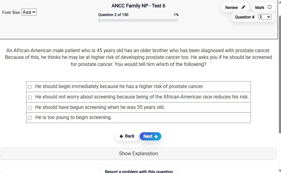
Answering a QuestionMultiple-choice item view with navigation controls and progress tracker.
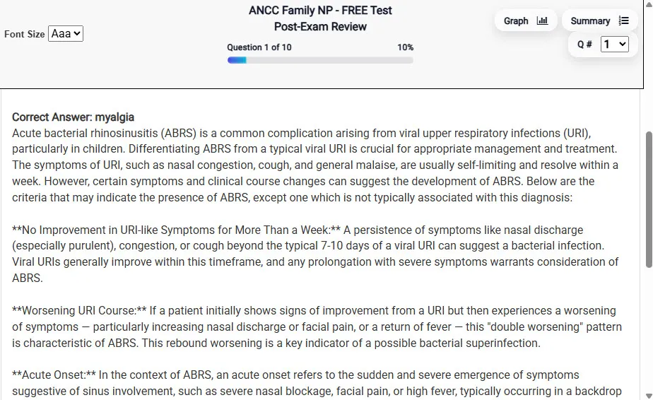
Detailed Explanation
Review mode showing chosen answer and rationale and references.
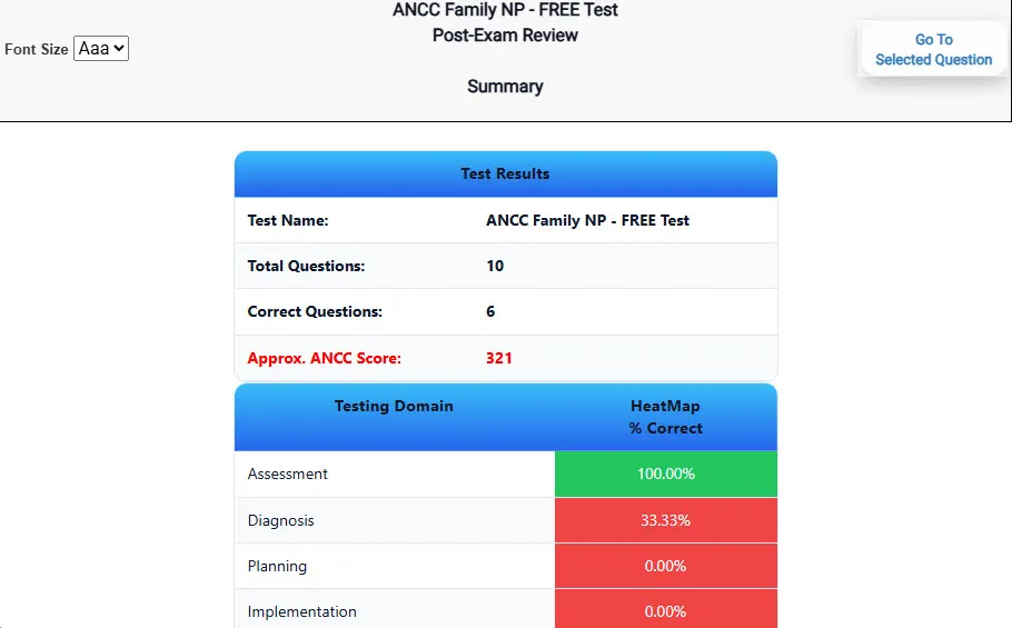
Review Summary 1
Summary with counts for correct/wrong/unanswered and not seen items.
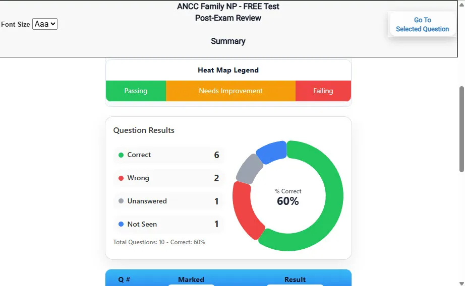
Review Summary 2
Advanced summary with category/domain breakdown and performance insights.
Screenshot
What Each Screen Shows
Answer Question Screen
Clean multiple-choice interface with progress bar.
Mark for review feature.
Matches real test pacing.
Detailed Explanation
Correct answer plus rationale.
Key concepts and guidelines highlighted.
Move between questions to fill knowledge gaps.
Review Summary 1
Overall results with total questions and scaled score.
Domain heatmap shows strengths and weaknesses.
Quick visual feedback on study priorities.
Review Summary 2
Chart of correct, wrong, unanswered, not seen.
Color-coded results for easy review.
Links back to missed items.
Top 10 Reasons to Use Exam Edge for your ABIM Cardiovascular Disease Exam Prep
Focused on the ABIM Cardiovascular Disease Exam
Our practice tests are built specifically for the ABIM Cardiovascular Disease exam — every question mirrors the real topics, format, and difficulty so you're studying exactly what matters.
Real Exam Simulation
We match the per-question time limits and pressure of the actual ABIM exam, so test day feels familiar and stress-free.
1 Full Practice Tests & 240 Unique Questions
You'll have more than enough material to master every ABIM Cardiovascular Disease concept — no repeats, no fluff.
Lower Cost Than a Retake
Ordering 5 practice exams costs less than retaking the ABIM Cardiovascular Disease exam after a failure. One low fee could save you both time and money.
Flexible Testing
Need to step away mid-exam? Pick up right where you left off — with your remaining time intact.
Instant Scoring & Feedback
See your raw score and an estimated ABIM Cardiovascular Disease score immediately after finishing each practice test.
Detailed Explanations for Every Question
Review correct and incorrect answers with clear, step-by-step explanations so you truly understand each topic.
Trusted & Accredited
We're fully accredited by the Better Business Bureau and uphold the highest standards of trust and transparency.
Web-Based & Always Available
No software to install. Access your ABIM Cardiovascular Disease practice exams 24/7 from any computer or mobile device.
Expert Support When You Need It
Need extra help? Our specialized tutors are highly qualified and ready to support your ABIM exam prep.
Pass the ABIM Cardiovascular Disease Exam with Realistic Practice Tests from Exam Edge
Preparing for your upcoming ABIM Cardiovascular Disease (CARDDIS) Certification Exam can feel overwhelming — but the right practice makes all the difference. Exam Edge gives you the tools, structure, and confidence to pass on your first try. Our online practice exams are built to match the real ABIM Cardiovascular Disease exam in content, format, and difficulty.
📝 1 ABIM Cardiovascular Disease Practice Tests: Access 1 full-length exams with 240 questions each, covering every major ABIM Cardiovascular Disease topic in depth.
⚡ Instant Online Access: Start practicing right away — no software, no waiting.
🧠 Step-by-Step Explanations: Understand the reasoning behind every correct answer so you can master ABIM Cardiovascular Disease exam concepts.
🔄 Retake Each Exam Up to 4 Times: Build knowledge through repetition and track your improvement over time.
🌐 Web-Based & Available 24/7: Study anywhere, anytime, on any device.
🧘 Boost Your Test-Day Confidence: Familiarity with the ABIM format reduces anxiety and helps you perform under pressure.
These ABIM Cardiovascular Disease practice exams are designed to simulate the real testing experience by matching question types, timing, and difficulty level. This approach helps you get comfortable not just with the exam content, but also with the testing environment, so you walk into your exam day focused and confident.




 General Exam Info
 Features
 Exam Details
 FREE Practice Test